MUTATION
It is a phenomenon which results in alteration of DNA sequences.
DNA
sequences alteration leads to changes in the genotype and the phenotype of an organism.
In addition to recombination, mutation is another phenomenon that leads to variation in DNA..
POINT MUTATION
Due to change in a single base pair of DNA!
| CODON | Mutant | Result |
|---|---|---|
| UUU | UUC | Silent Mutation |
| UUU | UUA | Mis-Sense Mutation |
| UGG | UGA | Non-Sense Mutation |
-
Addition/Deletion
of base pairs of DNA.- causes frame-shift mutations
- Frameshifts Since protein-coding DNA is divided into codons three bases long, insertions and deletions can alter a gene so that its message is no longer correctly parsed. These changes are called frameshifts.
-
Deletion/Duplication
of a segment of DNA- Result in alteration in chromosomes.
- Known as chromosomal abnormalities or aberrations.
- Chromosomal aberrations are commonly observed in cancer cells.
Mutagens
There are many chemical and physical factors that induce mutations. UV radiations can cause mutations in organisms – it is a mutagen.
Mendelian disorders are mainly determined by alteration or mutation in the single gene.
May be Dominant or recessive.

Sickle-cell anaemia is an autosome linked recessive
..trait that can be transmitted from parents to the offspring when both the partners are carrier for the gene (or heterozygous). The disease is controlled by a single pair of allele, HbA and HbS.
Sickle Cell Anemia
-
Genotype-1-HbA HbA.
Result : Normal Phenotype -
Genotype-2-HbA HbS.
Result : Apparently unaffected but they are carrier of the disease as there is 50 per cent probability of transmission of the mutant gene to the progeny, thus exhibiting Sickle Cell Trait -
Genotype-2-HbS HbS.
Diseased phenotype : Sickle Cell Anemia
Point MUTATION
-
GAG to GUG Substitution of
Glutamic acid (Glu) by Valine (Val) at the sixth position of the beta globin chain of the haemoglobin molecule. -
Mutant haemoglobin molecule
undergoes polymerisation under low oxygen tension causing the change in the shape of the RBC from biconcave disc to elongated sickle like structure.
Thalassemia.
Thalassemia is autosome-linked recessive blood disease. The defect could be due to either mutation or deletion which ultimately results in reduced rate of synthesis of one of the globin chains (α and β chains) that make up haemoglobin.
Alpha and Beta Thalassemia
The thalassemias are divided into two basic categories based on the affected chain. Therefore, alpha-thalassemias are due to defects in alpha-globin synthesis while beta-thalassemias are due to defects in beta-globin synthesis.
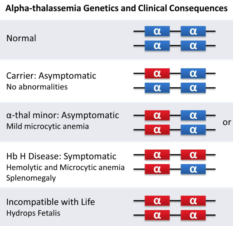
Alpha Thalassemia
Genes HBA1 and HBA2 on chromosome 16
- Alpha: Production of α globin chain is affected
Due to mutation or deletion of one or more of the four genesThe more genes affected, the less alpha globin molecules produced.
β Thalassemia
β Thalassemia is controlled by a single gene HBB on chromosome 11 of each parent and occurs due to mutation of one or both the genes.
Thalassemia Minor or Thalassemia Trait.
In this condition, the lack of beta protein is not great enough to cause problems in the normal functioning of the hemoglobin. As in mild alpha thalassemia, physicians often mistake the small red blood cells of the person with beta thalassemia minor as a sign of iron-deficiency anemia and incorrectly prescribe iron supplements.
Thalassemia Major or Cooley’s Anemia.
This is the most severe form of beta thalassemia in which the complete lack of beta protein in the hemoglobin causes a life-threatening anemia that requires regular blood transfusions. These extensive, lifelong blood transfusions lead to iron-overload which must be treated with chelation therapy to prevent early death from organ failure.
Thalassemia
v/s
SCA
Thalassemia is a quantitative problem of synthesising too few globin molecules while the Sickle-cell anaemia is a qualitative problem of synthesising an incorrectly functioning globin.
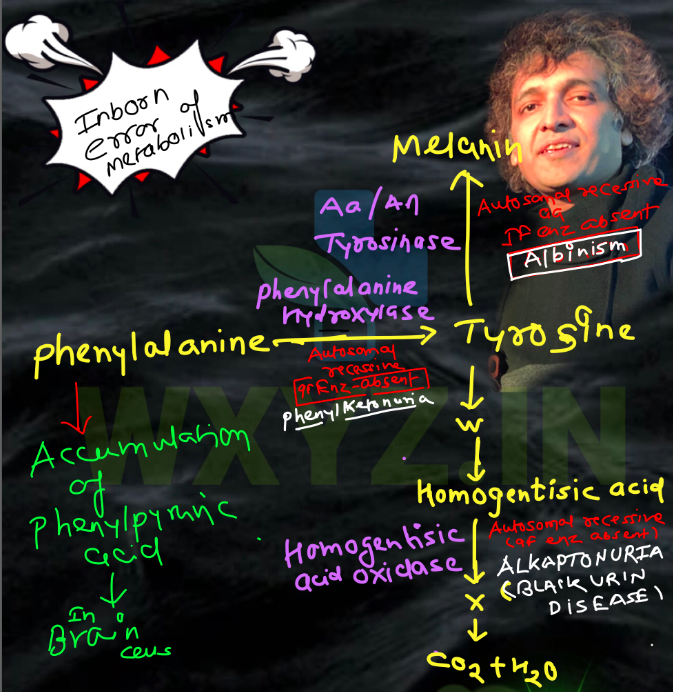
Inborn error of metabolism
by Archibald Garrod
- Albinism: Autosomal Recessive Disorder
- PKU:Autosomal Recessive Disorder
In Phenylketonuria : The affected individual lacks an enzyme that converts the amino acid phenylalanine into tyrosine. As a result of this phenylalanine is accumulated and converted into phenylpyruvic acid and other derivatives. Accumulation of these in brain results in mental retardation. These are also excreted through urine because of its poor absorption by kidney.
X-linked
Recessive trait shows transmission from carrier female to male progeny.
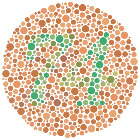
Colour Blidness
Sex(X)linked recessive disorder
Defect in either red or green cone of eye resulting in failure to discriminate between red and green colour.A daughter will be colour blind, when her mother is a carrier and her father is colour blind.
- Occurence: 8 per cent of males
- Occurence: 0.4 per cent of females
- Ishihara: normal People will see the number 74. People with color blindness will either see the number 21 or nothing.
Haemophilia
-
01. X linked recessive
a single protein (AHF-Anti Hemophilic Factor-VIII) that is a part of the cascade of proteins involved in the clotting of blood is affected.
-
02. Problem
In affected individual a simple cut will result in non-stop bleeding
-
03. Rare Possibility
a female becoming a haemophilic is extremely rare
-
Royal Disease
- Queen Victoria was a carrier of the disease.
- The family of Queen Victoria shows a number of haemophilic descendents
- Lack of Factor VIII
-
1.
Chromosomal disorders
caused due to absence or excess or abnormal arrangement of one or more chromosomes.
-
2.
Aneuploidy
Failure of segregation of chromatids during cell division cycle results in the gain or loss of a chromosome(s), called aneuploidy.
-
3.
Polyploidy
Failure of cytokinesis after telophase stage of cell division results in an increase in a whole set of chromosomes in an organism.This condition is often seen in plants.
Aneuploidy
-
Non Disjunction
During Meiosis (Failure of segregation of chromatids) -
2n+1 Trisomy
an additional copy of a chromosome may be included in an individua -
2n-1 Monosomy
an individual may lack one of any one pair of chromosomes. -
2n-2 Nullisomy
-
2n+1+1 Double Trisomy
Tetrasomy=2n+2 -
2n-1-1 Double Monosomy
...
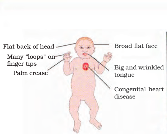
Down’s Syndrome
Trisomy of 21.
The affected individual is short statured with small round head, furrowed tongue and partially open mouth. Palm is broad with characteristic palm crease. Physical, psychomotor and mental development is retarded
- Described by: Langdon Down (1866)
- Occurence: 1/700
- Ask about: simian crease
- Klinefelter’s Syndrome 47, XXY
- Gynaecomastia
- Sterile
- Overall development Masculine
- Turner’s Syndrome 45, X0
- sterile
- Ovaries-Rudimentary
- Lack of other secondary sexual characters
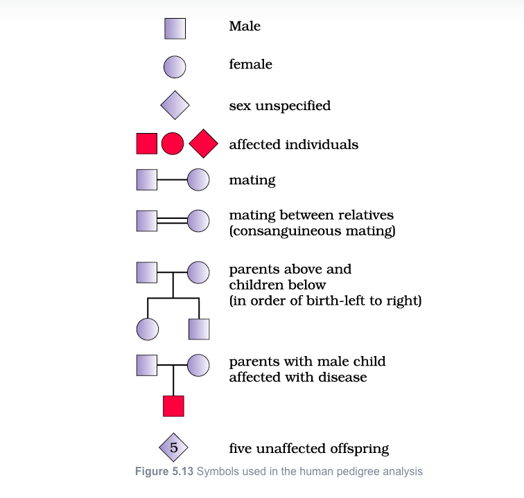
Pedigree analysis
family tree over generations.
Study of the family history about inheritance of a particular trait in humans, Since control crosses that can be performed in pea plant, are not possible in case of human beings.
- Method:analysis of traits in a several of generations of a family
- Strong tool:utilised to trace the inheritance of a specific trait, abnormality or disease.
YouTube Channel
Mendelism and Molecular Biology Lectures Available
Watch Complete Series on Youtube.com : YouTube Channel